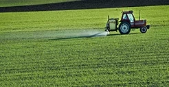
Residual Pesticides
Food and Beverages

Food industry is the largest industry of the global economy and it includes various sectors such as agriculture, manufacturing, production, processing, R&D, retail and more. It has global impacts and influences various other industry. Food is an essential part of our lives. With the current developments in the food industry, the demand, availability, access and trends of food have evolved to better cater to our needs and wants globally. Above all, the safety and quality of food is key to provide the world with food that is safe and nutritious.
-
-
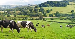
Veterinary Drugs
-
Food Additives
-
Beverage
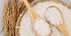
Brown Rice
Formula Milk Powder and Dairy Milk
Supplements
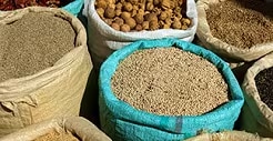
Other Foods and Seasonings
-
Natural Toxins
-
Extractable and Leachable
-
Edible Oils
-
PFAS in Food
Microorganism
Environmental pollutant
Substances formed in cooking or spoilage
Foreign substances
-
Aroma Analysis
-
Vitamins
Minerals
Fatty acids
Flavonoids
Carotenoids
Amino acids
Organic Acids
Sugar
Sweetener
Bitterness
-
Beverage
Food
-
Halal
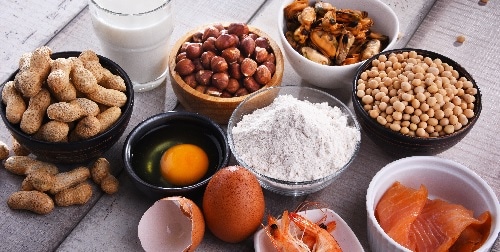
Food Allergen
-
Texture
-
Color


Featured Applications