Benchtop MALDI-TOF Imaging Starter Kit - Applications
Applications
| Applications | Date Creation Date |
|---|---|
2023-01-25 | |
2023-01-25 | |
2023-01-25 |

Most of the documents on the LITERATURE is available in PDF format. You will need Adobe Acrobat Reader to open and read PDF documents. If you do not already have Acrobat Reader, you can download it free at the Adobe's Website. Click the GET ADOBE READER icon on the left to download a free copy of Adobe Acrobat Reader.
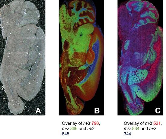
Analysis of lipids in full rat brain with 30 µm and 50 µm spacing

MALDI images of full rat brain using FlexiVision-mini ITO glass slide. A, optical scan; B, positive ion mode image from MALDI-8020 using 50 µm spacing; C, negative ion mode image from MALDI-8030 using 30 µm spacing.
Sample B
| Matrix | DHB, sublimated with Shimadzu iMLayer device |
|---|---|
| Measurement region | 67,024 pixels |
| Measurement time | around 1.9 hours |
| Experiment details | Laser repetition rate of 200 Hz and 20 shots per pixel |
Sample C
| Matrix | 9-AA, sublimated with Shimadzu iMLayer device |
|---|---|
| Measurement region | 275,210 pixels |
| Measurement time | around 3.8 hours |
| Experiment details | Laser repetition rate of 200 Hz and 10 shots per pixel |
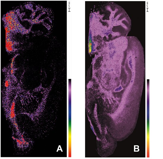
Large molecule imaging (protein/ on-tissue digestion) with 50 µm spacing

MALDI images of protein and peptides in full rat brain using FlexiVision-mini ITO glass slide and MALDI-8020. A, MBP protein, (m/z 14124); B, MBP peptide (HGFLPR, m/z 726.39)
Sample A
| Matrix | Sinapinic acid, sprayed with Shimadzu iMLayer AERO device |
|---|---|
| Measurement region | 68,836 pixels |
| Measurement time | around 3.8 hours |
| Experiment details | Laser repetition rate of 100 Hz and 20 shots per pixel |
Sample B
| Matrix | CHCA, sprayed with Shimadzu iMLayer AERO device |
|---|---|
| Measurement region | 67,482 pixels |
| Measurement time | around 2.8 hours |
| Experiment details | Laser repetition rate of 200 Hz and 30 shots per pixel |


