Measurement of the Physical Properties of HeLa Cells
The shape and elastic modulus of HeLa cells in a culture solution were measured using a scanning probe microscope.
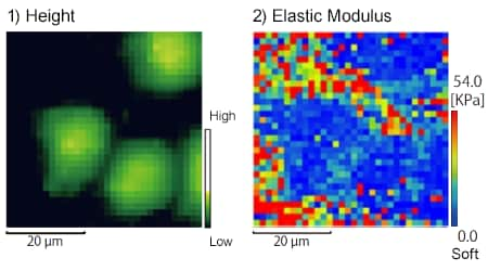
 Fig. 1 Mapping of the Shape and Elastic Modulus
These are the mapping results for the shape and elastic modulus (Fig. 1).
1) This shows the height information. The parts of the incubator surface without cells appear black, and the parts with cells are colored.
2) This shows the elastic modulus. The soft parts are blue, and as the hardness increases, the color changes to red.
The relationship between the elastic modulus (softness) of the cellular part and the cell height was evident.

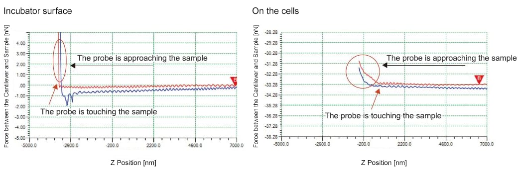
Fig. 2 Force Curve
The force curves for the incubator surface and the cell surface were compared (Fig. 2).
It is evident that after the probe makes contact with the sample, the curve rises more gently for the cells in comparison to the incubator surface. This indicates that the cells are softer, so it is possible to obtain quantitative data by analyzing the force curve.
(Data provided by: Professor Hirotaka James Okano, Research Associate Chikako Hara, Division of Regenerative Medicine, The Jikei University School of Medicine)


